|
Complications with Barrett's
Esophagus *
Patient B: 42 year old male with severe heartburn. Endoscopy showed
HH with severe exudative esophagitis. The patient was treated with high dose
Omeprazole.
Several months later endoscopy showed a 5 cm columnar mucosa. Biopsy showed
high grade dysplasia as well as invasive carcinoma.
1. High grade dysplasia
in Barrett’s
esophagus
(high power):
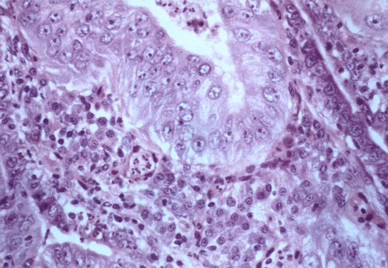
Figure 1
The high grade dysplasia is recognized
by markedly atypical cells with large pleomorphic nuclei with prominent nucleoli
and
a loss of polarity within the
epithelium. The epithelium is delimited by a basement membrane and surrounded
by inflammatory cells, mostly plasma cells with a few lymphocytes and pmn’s.
2. Invasive adenocarcinoma in association with high grade dysplasia
in Barrett’s
esophagus (high power):
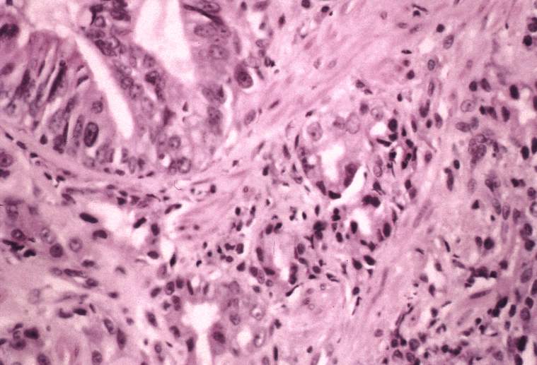
Figure 2
Small round glandular structures with atypical nuclei are present in an infiltrative
pattern in a reactive fibrous stroma. High grade dysplasia is in the epithelium
enclosed by the basement membrane in the upper left corner.
The following narrative provides more information on the relationship between
Esophageal Adenocarcinoma and GERD: Epidemiology of Adenocarcinoma
* Pathology slides of our patients were provided by Dr. James P. Kolton of
Caritas Norwood Hospital, Norwood, MA.
|